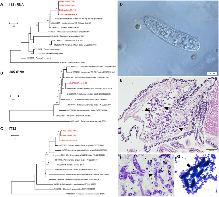
Fig. 2. Phylogenetic and microscopic analyses of Philaster sp. in abnormal D. antillarum.
(A to C) Phylogenetic representation of 18S rRNA and 28S rRNA Philaster-like sequences recovered from transcriptomes and 18S rRNA and ITS2 amplified from cultures FWC1 and FWC2 (cultured from abnormal urchins from an affected patch reef off Key Largo, Florida) and USF152 (cultured from an affected urchin during the experimental challenge study).These sequences are compared to homologous sequences of related species obtained from GenBank (accession numbers precede species names). Trees were constructed by maximum likelihood, the Jukes-Cantor substitution model, and with uniform rates of substitution. Bootstrap values of >50 (from 100 replicates) are indicated at nodes. Ciliates with morphologies similar to described Philaster sp. were observed by wet mount light microscopy (Nomarski prism) (D), in the base portion of the spine shaft by histopathology [indicated by arrowheads in (E) hematoxylin and eosin and (F) thionin], and in stained coelomic fluid cytology [indicated by arrowheads in (G); DiffQuick]. Photo credits: (D) to (F) Y. Kiryu and (G) T. Work.

