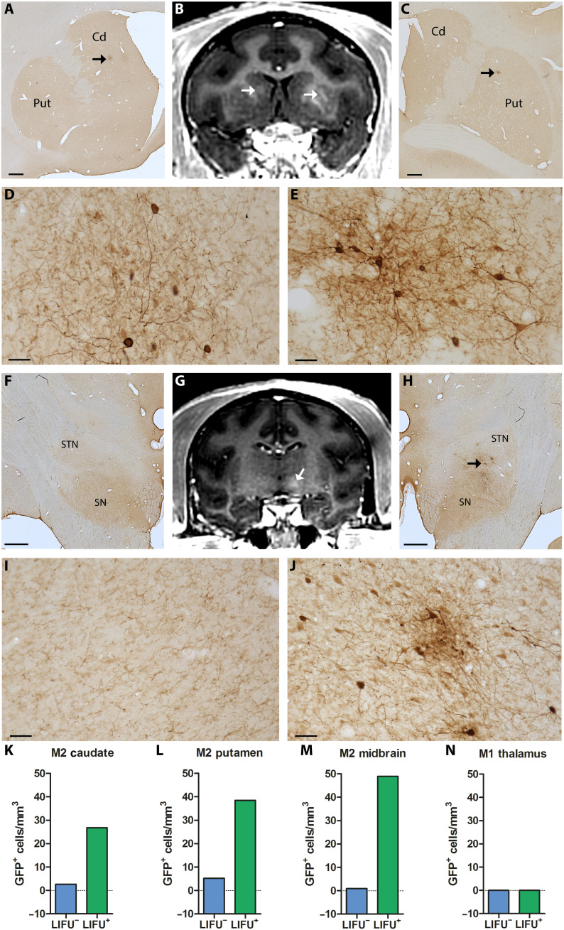
Fig. 4. Multitargeted focal delivery of modified AAV9 vector in the striatum and midbrain.
In M2, the distribution areas of GFP+ cells in the caudate nucleus [A, low magnification; D, higher magnification taken from the site specified by the arrow in (A)] and the putamen [C, low magnification; E, higher magnification taken from the site specified by the arrow in (C)] overlap the regions of Gd extravasation observed immediately after sonication (arrows in B). Similarly, GFP+ cells in the midbrain [H, low magnification; J, higher magnification taken from the site specified by the arrows in (H)] are distributed within the hyperintense BBB-opened area [arrow in (G)]. The same region on the contralateral side served as a negative control showing no hyperintense signal (G) or GFP expression (F and I, low and higher magnification, respectively). Number of GFP+ cells in the BBB-opened regions (LIFU+) and their contralateral non-opened regions (LIFU−) in the caudate nucleus (K), putamen (L), and midbrain (M) of M2 and in the thalamus of M1 (with NAbs) (N). Cd, caudate nucleus; Put, putamen; SN, substantia nigra; STN, subthalamic nucleus. Scale bars, 1 mm (A, C, F, and H) and 50 μm (D, E, I, and J).

