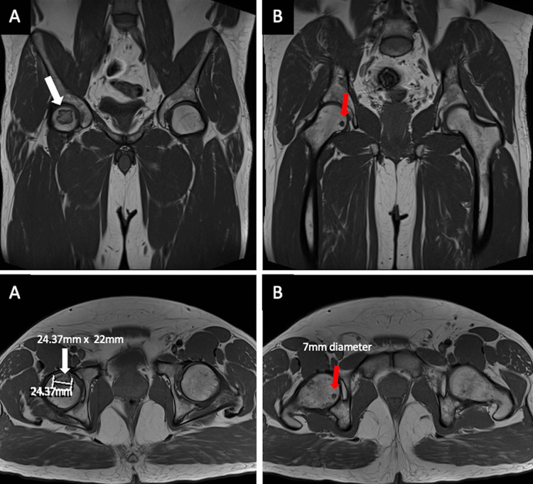
Figure 2. MRI radiographs demonstrating (A) a larger AVN lesion (white arrows), abutting the right anterior superior femoral cortex, measuring 25 x 22 mm, and (B) a smaller AVN lesion (red arrows), located more medially, measuring 7 mm in diameter. The right femoral head remains spherical in shape, with no articular irregularity, collapse, or surrounding tissue degeneration.
AVN: avascular necrosis.

