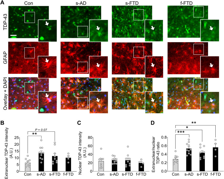
Fig. 1. Human astrocytes have increased extranuclear TDP-43 accumulation in AD and FTD.
(A) Representative images of TDP-43 immunoreactivity (green) in human postmortem hippocampal sections from nondementia controls (Con), sporadic AD (s-AD), sporadic FTD–TDP-43 (s-FTD), or familial FTD–TDP-43 (f-FTD) cases. The astrocyte marker GFAP (red) was used to visualize astrocytic cell bodies and main processes, and DAPI (blue) was used to visualize cell nuclei within individual astrocytes. Scale bar, 50 μm. (B to D) Quantification of TDP-43 immunoreactivity within different astrocytic subcellular regions. A.U., arbitrary units. One-way analysis of variance (ANOVA): F(3,28) = 4.21, P = 0.014 (B); F(3,28) = 0.34, P = 0.80 (C); and F(3,28) = 7.56, P = 0.0007 (D); Dunnett’s post hoc test: *P < 0.05, **P < 0.01, and ***P < 0.001 versus controls.

