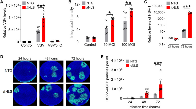
Fig. 6. Astrocytic TDP-43 affects antiviral defenses in a cell-autonomous manner.
(A) Primary astrocytes from NTG and ΔNLS mice were infected with VSV [100 multiplicity of infection (MOI)] for 24 hours. Some wells were also transfected with poly(I:C) (pI:C). VSV levels were measured by RT-qPCR. Two-way ANOVA: F(2,41) = 34.64, P < 0.0001 for interaction and F(1,41) = 40.60, P < 0.0001 for genotype. Bonferroni post hoc test: ***P < 0.001 versus NTG. (B) Primary astrocytes were infected with adenovirus tagged with enhanced green fluorescent protein (eGFP) at indicated MOIs for 24 hours. eGFP levels were measured by quantitative microscopy. F(2,24) = 3.47, P = 0.047 for interaction and F(1,24) = 13.73, P = 0.0011 for genotype. Bonferroni post hoc test: *P < 0.05 and **P < 0.001 versus NTG. (C) Primary astrocytes were infected with HSV-1 tagged with eGFP (0.01 MOI) for 24 or 72 hours. Glycoprotein B (gB) DNA levels were normalized to 18S DNA per sample. Two-way ANOVA: F(1,12) = 15.66, P = 0.0019 for genotype and F(2,12) = 9.225, P = 0.0037 for interaction. Bonferroni’s post hoc test: ***P = 0.0003 versus NTG for 72 hours. (D and E) Conditioned medium was collected from primary NTG or ΔNLS astrocytes after infection with HSV-1–eGFP (0.01 MOI). Astrocytes were washed 3 hours after infection, and conditioned medium was analyzed after indicated durations using the plaque assay in Vero cells. (D) Representative images of Vero cells after treatment with conditioned medium from NTG or ΔNLS astrocytes that were infected with HSV-1–eGFP for indicated durations. Scale bar, 1200 μm. (E) Viral particles in conditioned medium from HSV-1–infected NTG or ΔNLS astrocytes. Two-way ANOVA: F(1,33) = 15.24, P = 0.0004 for genotype and F(2,33) = 5.58, P = 0.0082 for interaction. Bonferroni’s post hoc test: ***P = 0.0009 versus NTG for 72 hours.

