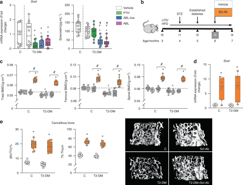
Fig. 5.
PTH and ABL decreased Sost expression in T2-DM bone, and the anti-sclerostin antibody restored and further increased BMD and cancellous bone in T2-DM. a Expression of the osteocytic marker Sost in bone and Sclerostin levels in serum of DM mice after treatment with PTH or ABL (at t4). b Study design: Male C57BL/6 DM mice as well as C mice were administered with vehicle or Scl-Ab once a week for 4 weeks. c Total, femoral and spinal BMD before treatment (at t3, grey bars) and after treatment (at t4) with vehicle (white bars) or Scl-Ab (orange bars). d Expression of Sost in bones of C and DM mice after (at t4) treatment with vehicle or Scl-Ab. e Micro-CT analysis of femur cancellous bone: BV/TV and trabecular thickness (Tb.Th), after treatment with vehicle or Scl-Ab and representative micro-CT images. n = 10-12 mice/group. Data are presented as box & whisker plots where each dot represents a mouse. ^P < 0.05 versus C mice by one-way ANOVA for (a) or two-way ANOVA with post hoc Dunnet’s correction for (c). For (a), *P < 0.05 versus T2-DM mice treated with vehicle and ϮP < 0.05 versus T2-DM mice treated with PTH by one-way ANOVA with post hoc Tukey’s correction. For (c–e), *P < 0.05 versus respective vehicle-treated mice by two-way ANOVA with post hoc Bonferroni’s correction. #P < 0.05 versus respective mice at t3 (grey bars) by one-way ANOVA with post hoc Tukey’s correction

