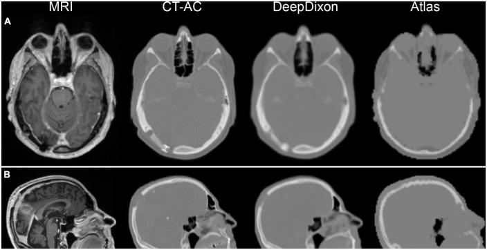
FIGURE 1.
Examples of magnetic resonance imaging (MRI) and corresponding attenuation maps for two post-surgery patients. First patient (A) has titanium alloy insert, which shows up has a thickening of tissue on DeepDixon, and not at all in the atlas method. Second patient (B) had part of the skull removed in the parietal region, which is well represented by DeepDixon. Particularly the facial and nasal regions are challenged in the atlas method leading to quantitative errors in brain directly bordering the skull base, anterior and posterior fossa, inferior temporal lobes, mesencephalon, pons, and cerebellum.

