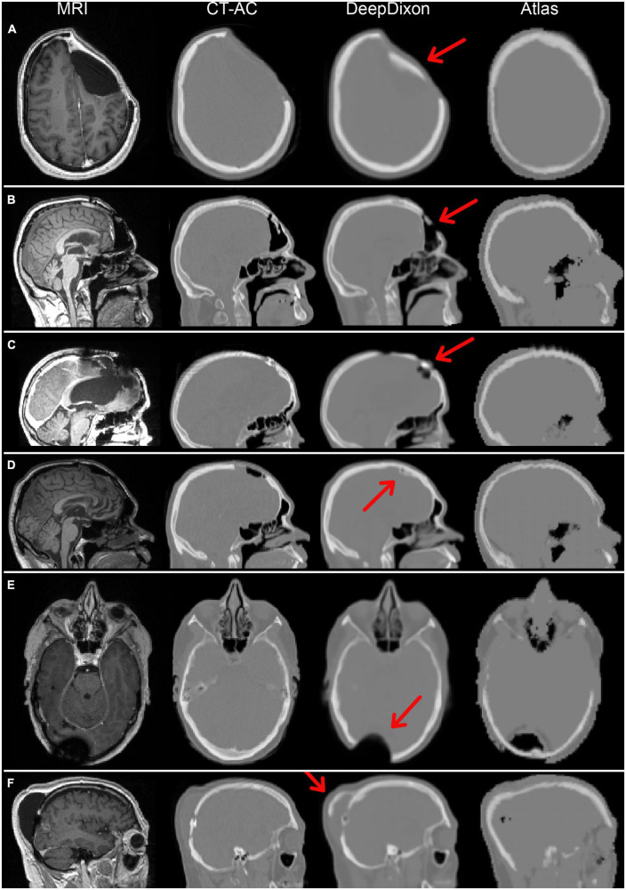
FIGURE 2.
Magnetic resonance imaging (MRI) and attenuation maps for all six patients with DeepDixon artifacts categorized as major. Two patients had follow-up scans, both with consistent artifacts, resulting in a total of 8/318 scans with major artifacts. The patients in (A–C,E) had biological tumor volume (BTV) > 1 ml, and were therefore included in the positron emission tomography (PET) evaluation. The primary errors were caused by false bone formation along titanium implant/soft tissue (A,D,F), or directly on brain in postoperative pneumocephalus (B), and metal associated signal voids (C,E).

