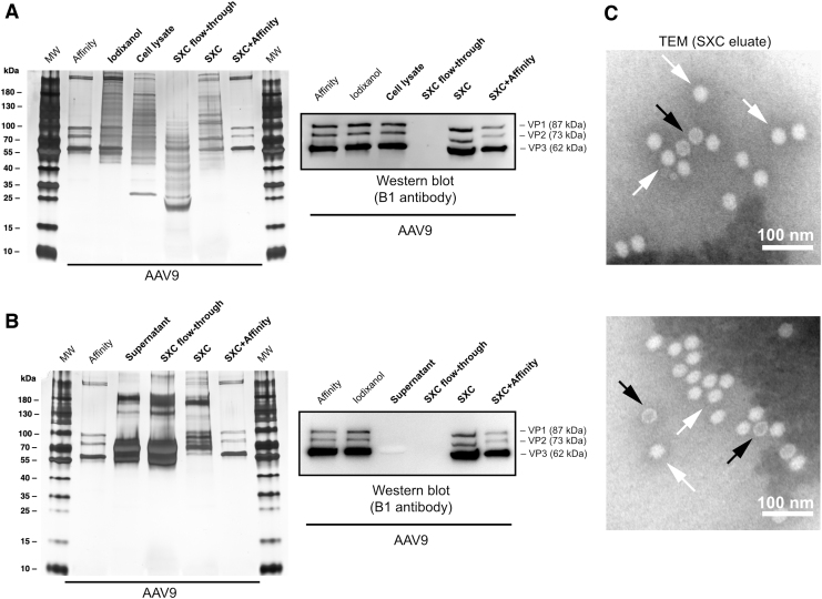
Figure 5.
Silver-stained SDS-PAGE and WB of SXC purifications from (A) cell lysates and (B) cell supernatants of AAV9. Affinity- and iodixanol-purified samples were included as reference. MW is the molecular weight marker. The viral proteins VP1, VP2, and VP3 are indicated. (C) TEM of SXC eluates. The AAV particles are homogeneous in shape and size with an approximate diameter of 25 nm. Genome-containing particles (white arrows) appear white in the negative staining, as opposed to empty capsids (dark arrows), which appear as a white rim with a dark core. Scale bars represent 100 nm. SDS-PAGE, sodium dodecyl sulfate–polyacrylamide gel electrophoresis; TEM, transmission electron microscopy; WB, Western blot.

