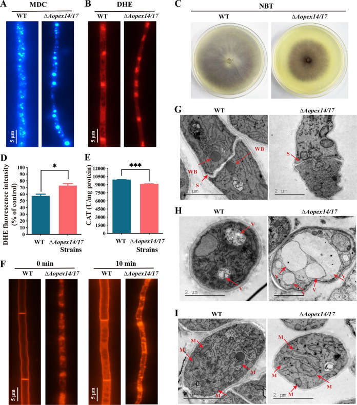
FIG 5.
Observation of intracellular autophagosomes, ROS accumulation, endocytosis, Woronin bodies, and vacuoles between the WT and ΔAopex14/17 mutant strains. (A) The autophagosomes of the WT and ΔAopex14/17 mutant strains were stained with monodansylcadaverine (MDC). Scale bar, 5 μm. (B) Representative images of dihydroethidium (DHE) staining to detect superoxide in the WT and ΔAopex14/17 mutant strains. Scale bar, 5 μm. (C) Nitroblue tetrazolium (NBT) staining for ROS production in mycelia of the WT and ΔAopex14/17 mutant strains. The dark color of the colony indicates increased ROS production. (D) Analysis of the DHE fluorescence intensity of the same weight of mycelium in the WT and ΔAopex14/17 mutant strains via multimode microplate reader. (E) Catalase (CAT) activity assay of the same weight of mycelium in the WT and ΔAopex14/17 mutant strains via multimode microplate reader. (F) FM4-64 staining to observe endocytosis in the WT and ΔAopex14/17 mutant strains at different times. Scale bar, 5 μm. (G to I) The WT and ΔAopex14/17 mutant strains were observed by transmission electron microscopy. WB, Woronin body; S, septum; V, vacuoles; M, mitochondrion. Scale bar, 2 μm.

