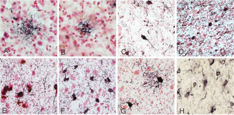
Figure 1.
Photomicrographs depicting PSP pathology. Tufted astrocytes captured on Gallyas silver stain in the middle frontal cortex (A) and in the putamen (B). Immunohistochemical staining for hyperphosphorylated tau (AT8 antibody) showing neuronal tangles in the globus pallidus (C), the subthalamic nucleus (D), the substantia nigra (E), basal pons (F), the dentate nucleus of the cerebellum (G), and coiled bodies in oligodendrocytes in the globus pallidus (H). Positive immunostaining is black and Neutral Red is used as a counterstain. Photomicrographs were taken at 40× magnification (A and B, H) and 20× magnification (C–H).

