Figure 2.
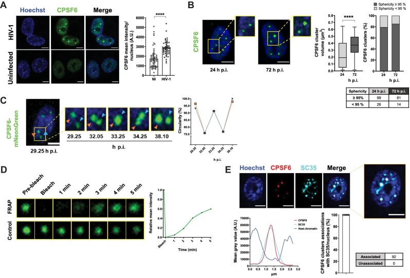
Phase-separation properties of HIV-1 MLOs. (A) Confocal microscopy images of THP-1 cells infected with HIV-1 (MOI = 5, 24 h p.i.) compared to uninfected cells. On the right, mean intensity of CPSF6 per nucleus ± SD (number of cells in the NI group = 49, number of cells in the HIV-1-infected group = 55). Unpaired t-test, ****P ≤ 0.0001. Two independent experiments were performed. (B) THP-1 cells were infected with HIV-1 (MOI = 5) and treated with NEV (10 µM). Confocal microscopy images of these cells at different time post-infection were compared (24 h p.i. vs. 72 h p.i). On the right, box plots of the volume (median: ∼0.19 vs. ∼0.37 µm3) and percentages of CPSF6 clusters that show ≥95% or <95% sphericity value (number of cells: 125 (24 h p.i.), 95 (72 h p.i)). Unpaired t-test, ****P ≤ 0.0001. Results of two independent experiments were analyzed from 3D acquisitions. (C) Frames extracted from a 10-h time-lapse microscopy in 2D (Supplementary Video S1) of THP-1 cells expressing CPSF6 mNeonGreen infected with HIV-1 (MOI = 10). The graph shows the circularity (normalized ratio between the contour and interior expressed as a percentage) along the MLO fusion–fission event. Representative of six independent experiments. (D) Frames extracted from a FRAP time-lapse microscopy (Supplementary Video S2) in THP-1 cells expressing CPSF6 mNeonGreen infected with HIV-1 (MOI = 10, 3 days p.i.). Representative of three independent experiments. The graph shows the recovery of the signal curve. Pre-bleach signal is set to 1 and bleach signal is set to 0. (E) Confocal microscopy images of THP-1 cells infected with HIV-1 (MOI = 10, 3 days p.i.). On the bottom, graphs show the intensity profile of Hoechst, CPSF6, and SC35 signals along the segment crossing the MLO (yellow in top right image) and the percentage of CPSF6 colocalizing with SC35 per nucleus ± SD. Results of two independent experiments were analyzed from 3D acquisitions. Scale bar, 5 µm.

