
Figure 4.
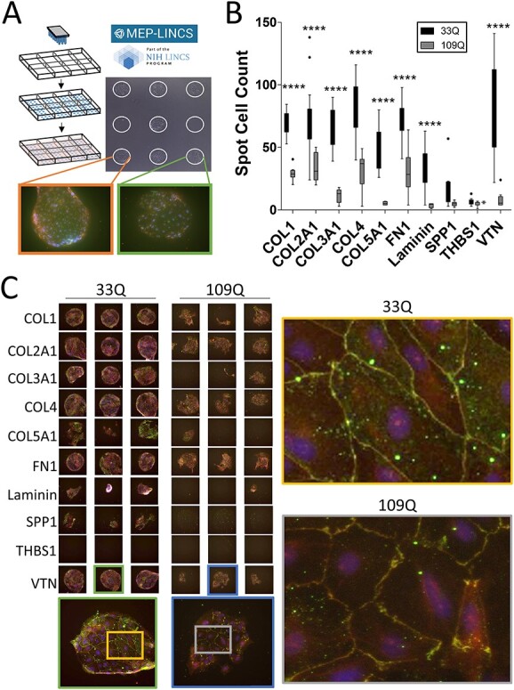
HD iBMECs have reduced adhesion and morphological deficits on ITG ligands. (A) MEMA schematic. Each spot is ~300 μm and is a unique ECM substrate plated at random on the array in multiple technical replicates. iBMECs are seeded, fixed and stained. Certain spots prevent TJ formation (green box), whereas others enabled TJ formation (orange box). (B) 33Q and 109Q iBMECs were assessed for adhesion by counting the number of Hoechst-positive cells per spot. All ITG ligands from the MEMA are shown. Two-way ANOVA with Bonferroni post-hoc. N = 1, n ≥ 11. (C) iBMECs stained for TJ proteins CLDN5 (green) and ZO1 (red) with a DAPI counterstain. Three representative technical replicates are shown. All technical replicates can be found in Supplementary Material, Figure S2. All ITG ligands shown (left). Morphological disruption of TJs is shown on VTN (right). N = 1, n ≥ 11. Each spot is ~300 μm.