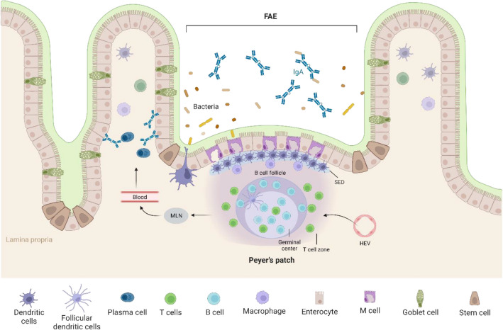
Fig. 1.
Schematic illustration of cellular composition of Peyer’s patches (PPs). PP follicles are enclosed by follicle-associated endothelium (FAE) containing M cells that shuttle luminal antigen into the PP. Subepithelial dome (SED) below the FAE contains high density of antigen-presenting cells. Lymphocytes enter PP via high endothelial venules (HEV) and form large B cell follicles and small T cell zones. The interactions between B cells and T cells at the follicle-T cell zone lead to expansion and differentiation of B cells. The activated B cells form germinal center, generating IgA-secreting plasma cells. The generated effector cells leave the PPs through efferent lymphatics and enter the circulation via mesenteric lymph nodes (MLNs) [32]. They home to the intestinal lamina propria from the blood circulation and the lamina propria plasma cells produce dimeric IgA that are transported across the epithelium. The secretory-IgA (S-IgA) can interact with bacteria in the gut lumen

