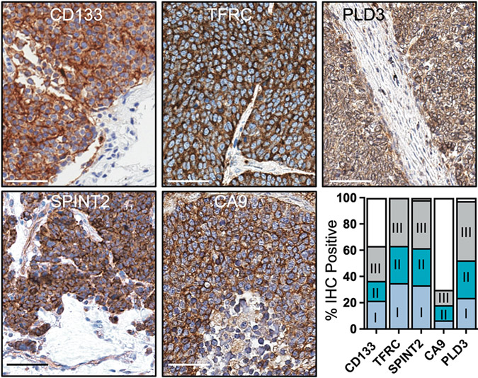
Figure 2. Antigens targeted by autoantibodies are upregulated in SCLC.
Representative immunohistochemical (IHC) images are shown of antigens targeted by peripheral autoantibodies on SCLC tissue microarrays (n=45 to 62 cases). Scale bar, 50μM. Bar graph contains positive or negative scoring of tissue microarray cores. Positive scores are further categorized by stage at diagnosis.

