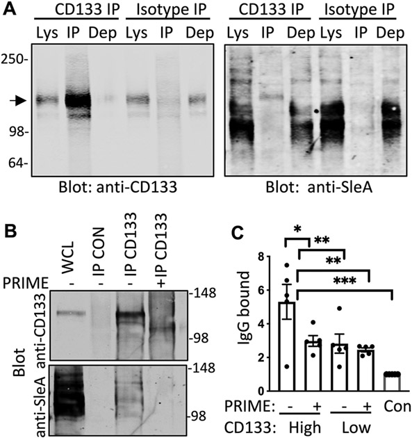
Figure 3. SCLC AAbs to CD133 target glycosylation motifs.
(A) Shown are immunoblots from H82 cell lysates (Lys) immunoprecipitated (IP) with CD133 antibody or isotype control and probed with anti-CD133 and anti-sLeA antibodies (N=3). Dep is depleted cell lysate after immunoprecipitation. The arrow indicates the expected protein size of CD133 at 133kDa (B) Shown are immunoblots from H82 lysates that were immunoprecipitated with isotype control antibody (CON) or CD133 antibody (N=3). Whole cell lysate (WCL) prior to immunoprecipitation is included to indicate the starting material. Lysates immunoprecipitated with CD133 antibody were then treated according to the manufacturer recommended deglycosylation protocol either with PRIME deglycosylase (+) or without enzyme (−). Immunoblots were probed with anti-CD133 and anti-sLeA antibodies. (C) ELISA quantification of autoantibody concentrations is shown for SCLC patient plasma that bound to CD133 from H82 cell lysates that had or had not been pre-treated with PRIME deglycosylase (n=5 per group). Con, negative control. Data are presented as individual values, the bar represents the mean ± SEM. *p<0.05, **p<0.005, ***p<0.0005 by student’s t test.

