
Figure 1.
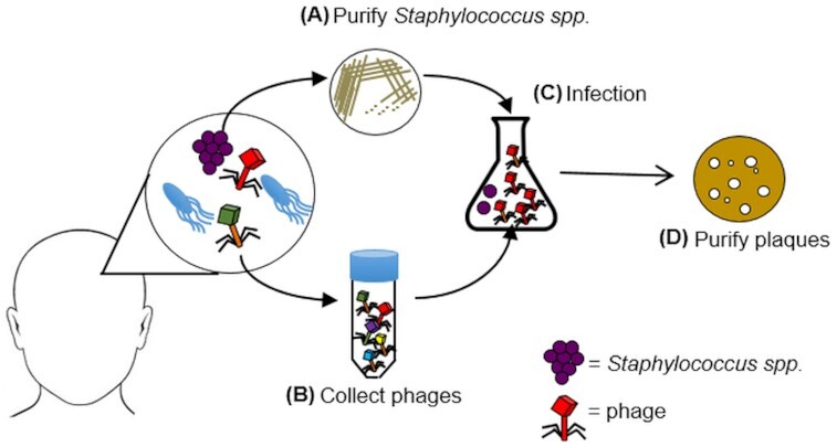
Schematic representation of the phage isolation method used in this study. Samples were taken from the skin of healthy volunteers. Commensal staphylococci were isolated using selective media then the species of each bacteria was determined via matrix-assisted laser desorption/ionization time-of-flight mass spectrometry (MALDI-TOF MS) (A). From the same skin site, a second sample was taken, filtered and phages were concentrated by centrifugation (B). S. epidermidis was then used as the host for phage propagation (C). Phage plaques were produced in double-layer agar plates for further purification and characterization (D).