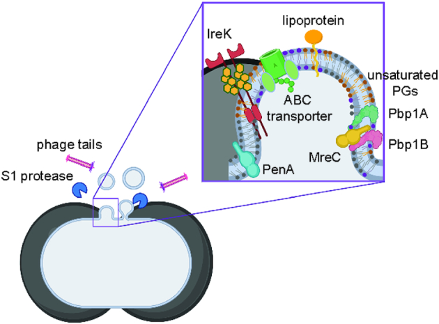
Figure 6.

Septal model for MV formation in E. faecalis. Cartoon depiction for MV formation from the septal region, where peptidoglycan is thinnest, during the cell divison. Enterococcus faecalisMVs are enriched in more flexible unsaturated PGs, and have a high abundance of septal proteins—Pbp1A, Pbp1B, MreC, PenA, that are involved in cell division, and IreK kinase, that is bound to un-crosslinked peptidoglycan through PASTA domains. Phage tails and S1 extracellular protease co-purify with MV fraction.
