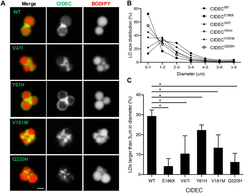
Fig 3. AMD CIDEC variants localize to lipid droplets (LDs) but cause a defect in LD enlargement.
(A) Representative images of GFP-tagged CIDEC wild-type (WT) or rare variants localized to LDs labeled in red by BODIPY 558/568. Scale bar: 2 μm. (B) Size distribution of LDs in pre-adipocytes expressing CIDEC WT or each of the rare variants (diameters in μm). (C) Percentage of LDs with a diameter larger than 3 μm. N = 3 (mean ± SD, Student’s t test, *p<0.05).

