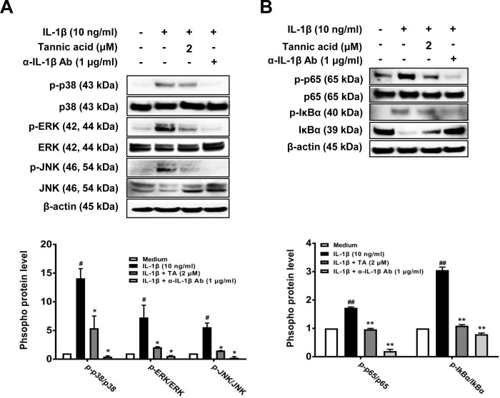
Fig 4. Effects of TA on IL-1β-induced MAPK and NF-κB activation in human OA chondrocytes.
Human articular chondrocytes from OA patients were seeded onto 6-well plates (3×10⁵ cells/well), and serum-starved cells were co-treated with various concentrations of TA (2 μM), or anti-IL-1 neutralizing antibody (1 μg/ml) and IL-1β (10 ng/ml) for 1 h (A) or 3 h (B). Protein expressions of phosphorylated and non-phosphorylated forms of p38, ERK, JNK, p65, and IκBα were detected by Western blot analysis. β-actin was used as a loading control. Band intensity was analyzed using Image J program. # p < 0.01 compared with medium only control group and * p < 0.01 compared with IL-1β-treated group.

