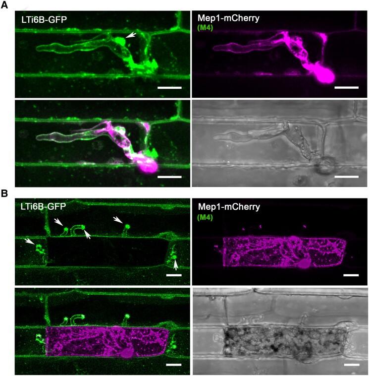
Figure 7.
The rice plasma membrane is invaginated and accumulates at the BIC during plant infection. Laser confocal micrographs of M. oryzae expressing Mep1-mCherry colonizing epidermal leaf cells of a transgenic rice line expressing plasma membrane-localized LTi6B-GFP. Images were captured at 24 hpi (A) and 36 hpi (B). The plant plasma membrane stays intact and invaginated at the early stages of plant infection. LTi6B fluorescence accumulates at the bright BIC, indicating the BIC is a plant membrane-rich structure. The fluorescence signal from secreted Mep1-mCherry is surrounded by the fluorescence signal of the rice cell plant plasma membrane marker LTi6B-GFP as the fungus invades new cells, but the initial epidermal cell is occupied and then loses viability, and Mep1-mCherry fluorescence fills the rice cell. Arrows indicate the BICs in the invaded neighboring cells. Scale bars = 10 um.

