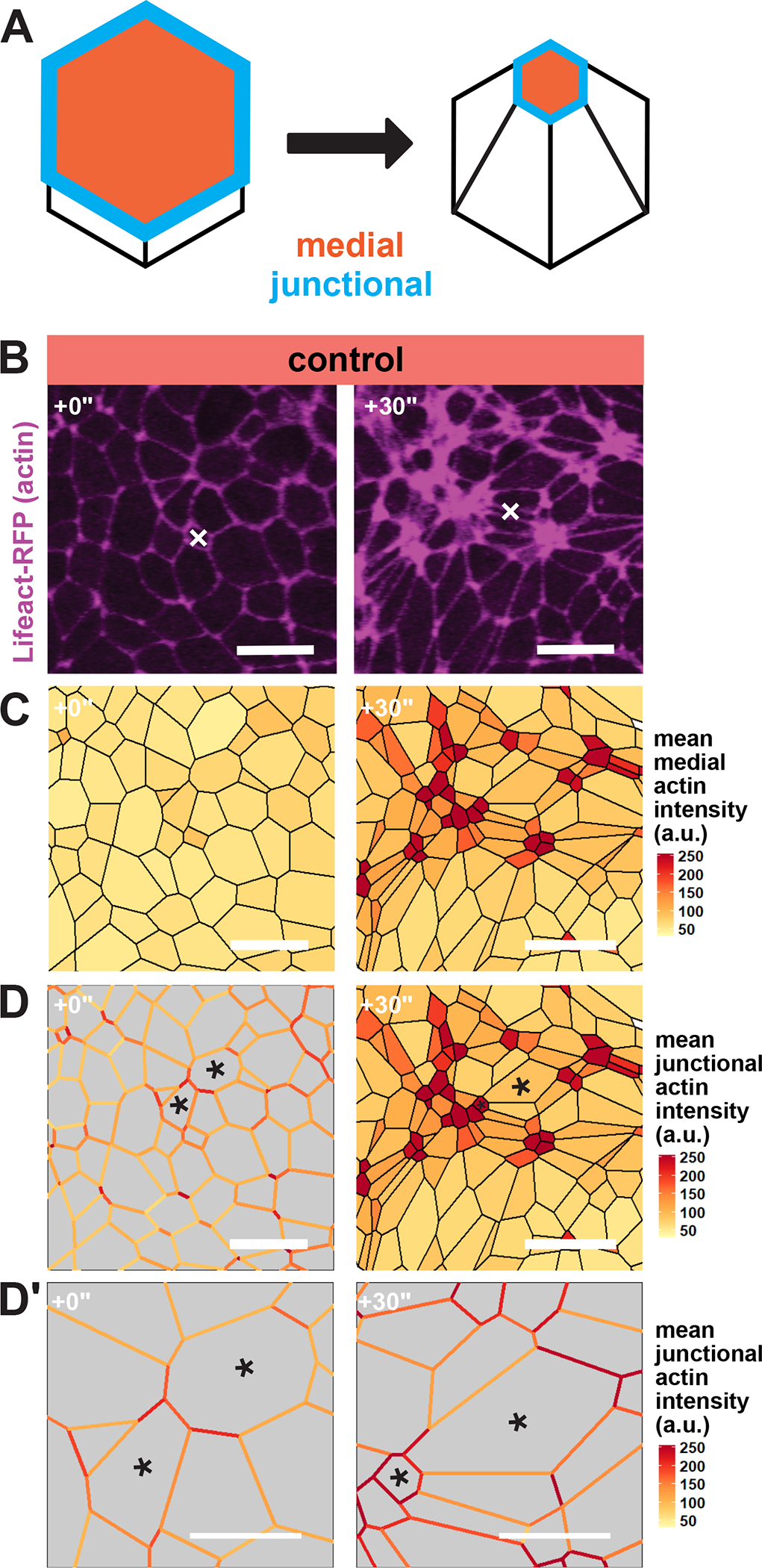
Figure 1:

Apical actin accumulation is heterogenous in the Xenopus neural ectoderm. A, Diagram of medial and junctional quantification domains on the apical surface of apically constricting cells. B, Lifeact-RFP localization in the anterior neural ectoderm of apically constricting cells. White “X” marks the same cell in each panel. Scale bar = 25μm. C, mean actin accumulation at the medial domain of neural ectoderm cells is heterogeneous. Actin intensity is measured in arbitrary units. Scale bar = 25μm. D, mean actin accumulation at individual cell-cell junctions is also heterogeneous. Actin intensity is measured in arbitrary units. Black asterisks mark the same cells in each frame. Scale bar = 25μm. D’, insets of cells from D. Scale bar = 12.5μm.
