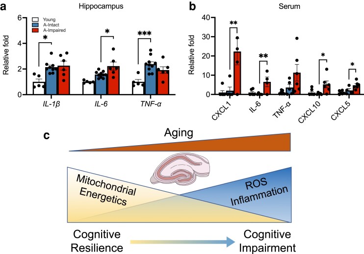
Fig. 5.
Increased inflammatory markers with impairment in aged mice. (a) Relative fold increases (compared to young, n = 5) in age-related hippocampal RNA expression of cytokines IL-1β (P = 0.017) and TNF-α (P = 0.0007) and the increase in IL-6 (P = 0.021) between aged–impaired (n = 9) and aged–intact (n = 6) groups. b) Bar plots depicting the changes in serum cytokine levels of CXCL1 (P = 0.0068), IL-6 (P = 0.0086), TNF-α, CXCL10 (P = 0.0199), and CXCL5 (P = 0.0426) between the aged–impaired (n = 5) and aged–intact (n = 5) groups relative to young (n = 6). c) Schematic representing mitochondrial mechanisms, oxidative stress, and inflammatory markers underlying cognitive heterogeneity. For all graphs, colors represent the following: young (white), aged–intact (blue), aged–impaired (red). Error bars depict the mean ± SEM. Significance was tested using one-way ANOVA (*P < 0.05; **P < 0.01; ***P < 0.001).

