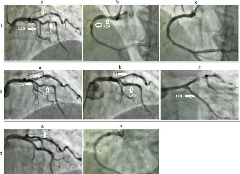
Figure 3.
Coronary angiography of the patient.
(1a) An 85% stenosis in the middle LAD (white arrow), an 85% stenosis in the proximity of thick D1 (white arrow), and a long 75 to 85% stenosis in the middle LCX (white arrow). (1b) A 90 to 95% stenosis in the proximal and middle RCA (white arrow). (1c) A DES was implanted in stenosis RCA. (2a) An 85% stenosis in the middle LAD (white arrow), an 85% stenosis in the proximity of thick D1 (white arrow), and a long 75 to 85% stenosis in the middle LCX (white arrow). (2b) An 85% stenosis in the proximity of thick D1 (white arrow) and a DES was implanted in stenosis of LAD. (2c) A DES was implanted in stenosis of LCX. (3a) A 70% stenosis in the proximity of thick D1. (3b) No stenosis in the RCA. D1 = first-diagonal branch, DES = drug-eluting stent, LAD = left anterior descending artery, LCX = left circumflex artery, RCA = right coronary artery.

