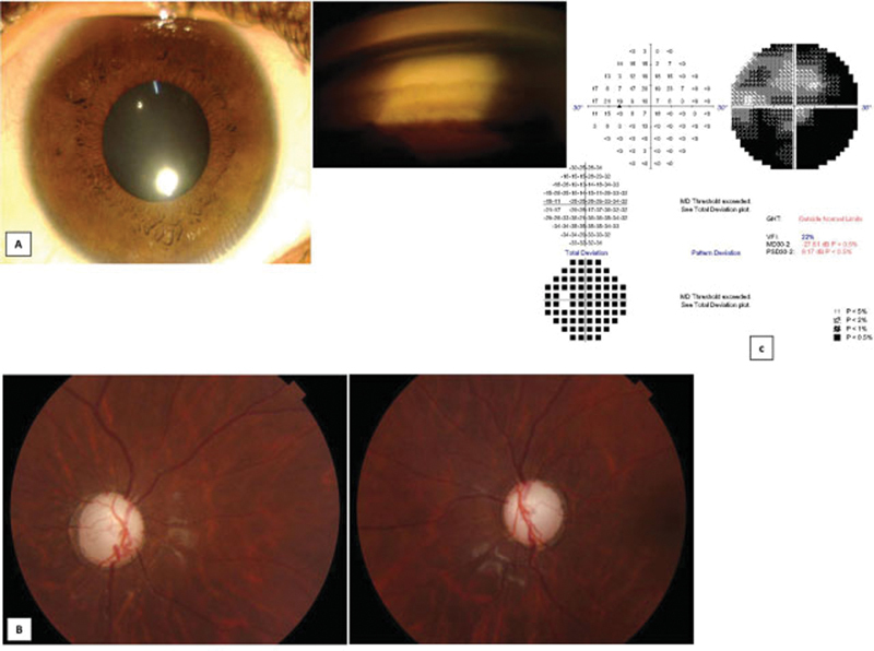
Fig. 2.

Anterior segment, optic disc photograph, and visual field of proband. ( A ) Anterior segment photograph of the proband along with goniophotograph showing wide open angles. ( B ) Optic disc photograph of the proband showing near total glaucomatous optic neuropathy in both eyes. ( C ) Visual field of the left eye of the proband showing severe visual field loss.
