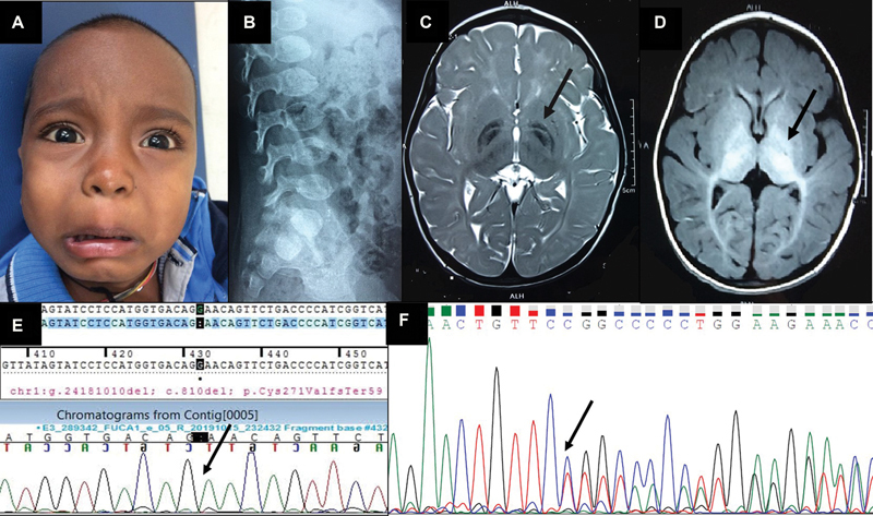
Fig. 1.

( A ) Close-up of the child's face showing the coarse facial features, mild ocular hypertelorism, and broad nasal bridge and thick lips. ( B ) Lateral radiograph of the lumbosacral spine showing the ovoid vertebrae and the anterior-inferior beaking indicative of dysostosis multiplex. ( C ) T2-weighted axial image of MRI brain showing bilaterally symmetrical hypointensity of both the globi pallidi with a central streak of hyperintensity between the medial and lateral segments of the globi pallidi, resembling the “eye-of-the-tiger” sign (pointed out by the arrow), along with symmetric hyperintensities in bilateral cerebral white matter. ( D ) T1-weighted axial image of MRI brain showing bilaterally symmetrical hyperintensity of globi pallidi, substantia nigra, and subthalamic nuclei (pointed out by the arrow). ( E ) Sanger sequence chromatogram of the proband showing the homozygous c.810del variant in the FUCA1 gene (the position of the single nucleotide deletion is marked with the arrow). ( F ) Sanger sequence chromatogram of the mother showing the heterozygous c.810del variant in the FUCA1 gene (the position of the single nucleotide deletion is marked with the arrow).
