Figure 3.

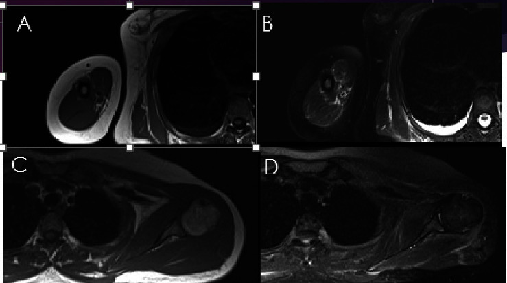
MRI of right arm T1W (a) and fat sat (b) and (c)-(d) MRI of left shoulder T1W (c) and fat sat (d) sequences. Note to the diffuse perimuscular and fascial edema more along the latissimus dorsi and rotator cuff muscles (arrows).

MRI of right arm T1W (a) and fat sat (b) and (c)-(d) MRI of left shoulder T1W (c) and fat sat (d) sequences. Note to the diffuse perimuscular and fascial edema more along the latissimus dorsi and rotator cuff muscles (arrows).