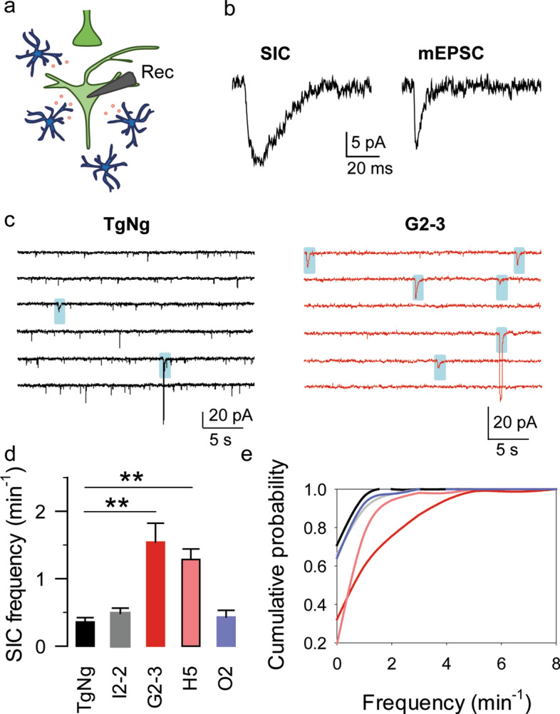
Fig. 4.

Astrocytic glutamate release is increased in G2-3 mice. a Scheme of the experimental approach. Note the glutamate (red) release by astrocytes (blue) and the recording CA1 pyramidal neuron (light green). b Representative SIC and mEPSC traces. c Representative SIC traces (shaded in blue) obtained from TgNg and G2-3 mice. d SICs per minute obtained from TgNg, I2-2, G2-3, H5 and O2 mice. Data are expressed as mean ± s.e.m. (**) p < 0.01. e Cumulative SIC frequency obtained from TgNg, I2-2 (the maximum difference between the cumulative distributions from TgNg and I2-2 mice, D, is 0.064902 with a corresponding p > 0.1), G2-3 (the maximum difference between the cumulative distributions from TgNg and G2-3 mice, D, is 0.386869 with a corresponding p > 0.1), H5 (the maximum difference between the cumulative distributions from TgNg and H5 mice, D, is 0.516783 with a corresponding p > 0.05) and O2 (the maximum difference between the cumulative distributions from TgNg and O2 mice, D, is 0.067116 with a corresponding p > 0.1) mice
