Fig. 6.
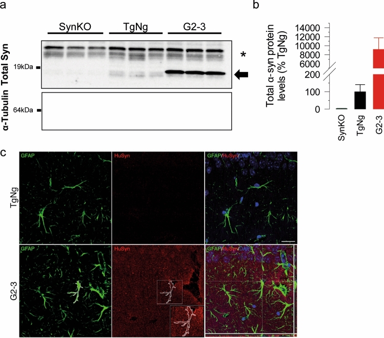
Analysis of α-synuclein expression in astrocytes. Lysates from primary astrocyte cultures from brains of SynKO, TgNg and G2-3 mice were analyzed for α-synuclein expression. a Immunoblot for α-synuclein from brain samples from SynKO, TgNg and G2-3 mice. α-tubulin is used as a loading control. Note that G2-3 astrocytes show strong α-syn reactivity (arrow). α-syn is present in TgNg astrocytes but not in SynKO astrocytes. *Non-specific band. b α-synuclein protein levels quantified from a. Data was normalized to α-tubulin levels and relativized to TgNg. c Astrocytes within the stratum radiatum of G2-3 mice, but not TgNg mice, contain intracellular human α-syntransgenic negative; TgNg glial fibrillary acidic protein, GFAP human alpha-synuclein, HuSyn. Scale bar = 25 μm

