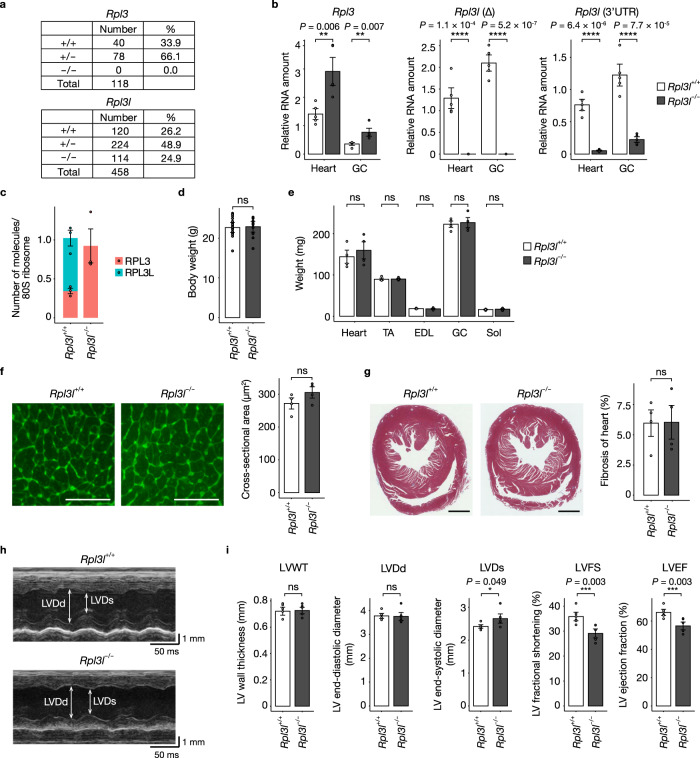
Fig. 2. RPL3L-deficient mice manifest impaired cardiac contractility.
a Number and percentage of mice of the indicated genotypes at weaning for the offspring produced by mating male and female heterozygotes for each mutation. b RT-qPCR analysis of Rpl3l and Rpl3 mRNA abundance in the heart and gastrocnemius (GC) of Rpl3l+/+ and Rpl3l−/− mice at 10 weeks of age (n = 5 mice). Rpl3l transcripts were measured with primers targeting either the deleted region (Δ) or the intact 3’UTR. c Number of RPL3 and RPL3L molecules per 80S ribosome as determined by MRM analysis of the polysome fraction from the heart of Rpl3l+/+ and Rpl3l−/− mice at 10 weeks of age (n = 3 mice). d, e Body weight of Rpl3l+/+ (n = 21 mice) and Rpl3l−/− (n = 12 mice) mice at 10 weeks of age and tissue weight for the heart as well as TA, EDL, GC, and Sol muscles of Rpl3l+/+ and Rpl3l−/− mice (n = 4 mice) at 18 to 19 weeks of age (e). f, g Representative images of WGA staining and Masson’s trichrome staining for heart sections from Rpl3l+/+ and Rpl3l−/− mice at 18 to 19 weeks of age (left panels) and quantitative analysis of muscle fiber size and the extent of fibrosis (blue staining) determined from such sections, respectively (n = 4 mice) (right panels). Scale bars, 50 μm (f) and 1 mm (g). h, i Representative echocardiographic images and quantitative analysis of LVWT, LVDd, LVDs, LVFS, and LVEF for Rpl3l+/+ and Rpl3l−/− mice at 18 to 19 weeks of age (n = 6 mice). All quantitative data in bar graphs are means ± s.d. *P < 0.05, **P < 0.01, ***P < 0.005, ****P < 0.001; ns not significant (unpaired two-tailed Student’s t test). Source data are provided as a Source Data file.

