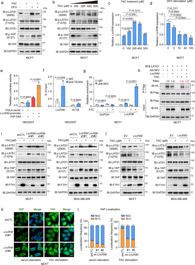
Fig. 4. The iron-triggered LncRIM-NF2 feedback loop hyperactivates YAP.
a Immunoblot was performed to examine the level of p-YAP (S127) and p-LATS1(S909, T1079). Serum-starved MCF-7 cells were treated with FAC (200 µM) or DFO (100 µM) for 24 h. b Serum-starved MFC7 cells were stimulated with different concentrations of FAC for 24 h. Immunoblot was performed to detect the level of p-YAP (S127) and p-LATS1(S909, T1079). c RT–qPCR detection of the LncRIM expression of MCF-7 cells stimulated with different concentration of FAC for 24 h. (mean ± SD, n = 3, one-way ANOVA analysis). d The expression of LncRIM in MCF-7 cells stimulated with different concentration of DFO for 24 h was analyzed with RT-qPCR. (mean ± SD, n = 3, one-way ANOVA analysis). e Luciferase reporter assay was performed of HEK-293T cells with overexpression of YAP-5SA and LncRIM promoter. (mean ± SD, n = 3, one-way ANOVA analysis). f YAP/TEAD4 directly regulates the transcription of LncRIM. CHIP-qPCR assay was performed by using IgG and TEAD4 antibodies. (mean ± SD, n = 3, two-sided Student’s t test). g RIP and RT–qPCR assays were performed to assess the interaction between LncRIM and NF2 of MCF-7 cells with or without FAC (200 µM) stimulation for 24 h. NF2 was immunoprecipitated by NF2 antibody and Protein A/G beads. IgG was used as the negative control. (mean ± SD, n = 3, One-way ANOVA analysis).(h) SFB-LATS1, HA-NF2 and LncRIM were co-transfected into MCF-7 cells with or without FAC (200 µM) stimulation for 24 h. SFB-LATS1 was immunoprecipitated by Flag beads. i, j Serum-starved control, LncRIM knockdown (i) or LncRIM overexpressed (j) MCF-7 and MDA-MB-468 cells were treated with FAC (200 µM) for 24 h, and the levels of p-LATS1(S909, T1079), p-YAP (S127) and FTH1 were detected by immunoblot. k, l Immunofluorescence staining of YAP in control and LncRIM knocked down serum-starved MCF-7 cells with or without FAC (200 μM) stimulation for 24 h (k). Scale bar, 10 µm. YAP localization in cells from three randomly selected fields of view was quantified (l). (mean ± SD, n = 3).

