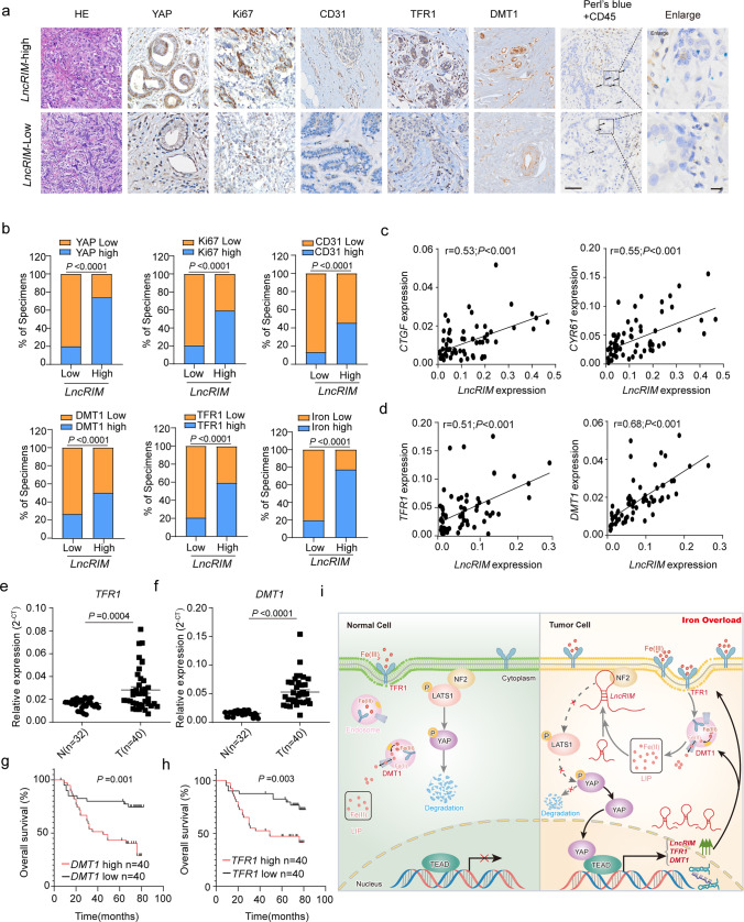
Fig. 6. High LncRIM expression correlates with poor clinical outcomes for breast cancer patients.
a The expression of LncRIM was positively correlated with the expression of YAP, Ki67, CD31, DMT1, and TFR1 in human breast cancer tissue (n = 98 including LncRIM-high and LncRIM-low subsets). Double staining of iron and CD45 in breast cancer cells (arrows). Scale bar, 100 µm. b Percentages of specimens with low and high LncRIM expression relative to the levels of YAP, Ki67, CD31, DMT1, TFR1, and iron (two-sided χ2 test). c The expression of LncRIM was positively correlated with CTGF and CYR61 as determined by two-sided chi-square test; R, correlation coefficient (n = 80 tumor patient samples). d The expression of LncRIM was positively correlated with DMT1 and TFR1 as determined by two-sided chi-square test; R, correlation coefficient (n = 80 tumor patient samples). e, f RT–qPCR detection of the DMT1 and TFR1 expression in tumor tissues (n = 40) and paired control tissues (n = 32). The horizontal black lines represent the median values. two-sided Student’s t test. g, h Recurrence-free survival analysis of the DMT1 status (g) and TFR1 status (h) of breast cancer patients (n = 80, Kaplan–Meier analysis with the Gehan–Breslow test). i Graphic illustration of the LncRIM-NF2-DMT1/TFR1 axis in cellular iron metabolism.

