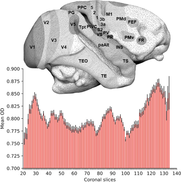
Fig. 5.

Mean Orientation Dispersion by slices N = 10. A right lateral view of a Saimiri brain surface (top, modified from Royo et al. 2021) is aligned to a histogram (bottom) representing the average orientation dispersion (OD) value for grey matter in each coronal slice. Error bars represent the standard error. FEF frontal eye field, FR frontal rostral field, INS insula, M1 primary motor area, PaAlt lateral parakoniocortex, PIVC parieto-insular vestibular cortex, PG inferior parietal lobule, PMv premotor area ventral, PMd premotor area dorsal, PPC posterior parietal cortex, PR presumptive parietal rostral, PV parietal ventral, S2 secondary somatosensory area, 1 2 3a 3b and 5 cytoarchitectonic areas 1 2 3a 3b and 5, TE temporal region, TEO temporo-occipital region, Tpt temporo-parietal region, TS temporalis superior cortex, V1 V2 V3 V4 and V5 visual areas 1 2 3 4 and 5, VS ventral somatosensory.
