Fig. 1.
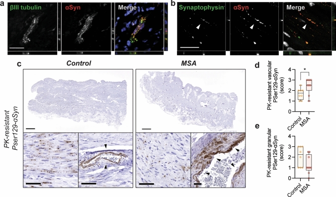
Detection of pathological ɑSyn in the urinary bladder of MSA patients and controls. Immunohistochemical analysis of endogenous ɑSyn in human urinary bladder shows ɑSyn expression in a βIII tubulin-positive intramural ganglia and b neuronal synapses (white arrows). For the detection of pathological ɑSyn urinary bladder tissue was treated with proteinase K (PK) and stained for PSer129- ɑSyn. c PK-resistant PSer129-ɑSyn is detected in urinary bladder tissue of both controls and MSA patients. A granular pattern of PK-resistant PSer129-ɑSyn is observed throughout the detrusor and lamina propria in addition to a vascular pattern where ɑSyn deposits around the vascular epithelium (black arrows) (scale bar represents 2 mm for overview figures and 50 µm for the detailed panels). The PSer129-⍺Syn immunostaining is localized to the perivascular area in the control subject whereas it extends more deeply into perivascular space for the MSA case. d Semi-quantitative analysis of PK-resistant PSer129-ɑSyn pathology in controls and cases shows significantly more vascular pathology for MSA cases (n = 8, *p = 0.02 with a non-parametric Mann–Whitney unpaired two-tailed t test) but e no differences in the granular PSer129-ɑSyn is detected (n = 8, p > 0.05 with a non-parametric Mann–Whitney unpaired two-tailed t test)

