Fig. 3.
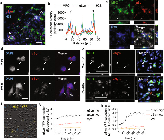
ɑSyn is found extracellularly in neutrophil extracellular traps. a Infected urinary bladders at 12 h show NETs with ɑSyn in the vicinity of red blood vessels in the lamina propria (scale bar is 15 µm, * and **indicate two blood vessels). b Intensity plot of MPO, H2B and ɑSyn along a 100 µm line in a shows overlap between the three markers. Highly ɑSyn expressing areas with strong intensity peaks are observed in NETs (open and closed arrows). c Confocal z-stacks of the NET in a (closed arrow) shows colocalization between MPO, ɑSyn and H2B (scale bar is 5 µm). d Primary human neutrophils express ɑSyn (white arrows), which is released via extracellular traps (black arrows) during infection with UPEC (10:1 MOI, scale bar is 20 µm). e In normal human urinary bladder neutrophils express relative low levels of ɑSyn while in a case of cystitis neutrophils with decondensing DNA release ɑSyn extracellularly (black arrows, scale bar is 40 µm). f The detection and release of extracellular DNA was visualized via live-imaging in ɑSyn-YFP-expressing dHL-60 stimulated with UPEC (10:1 MOI). Control cells are WT dHL-60 cells that do not express ɑSyn. g After infection control cells show background detection of fluorescent signal. ɑSyn-YFP low and high expressing cells show increasing fluorescent ɑSyn-YFP with h increased detection of ɑSyn-YFP in extracellular traps (experiment was performed in triplicate and showing one representative experiment)

