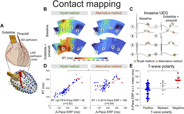
FIGURE 3.
RT determined by the Wyatt and alternative method through contact mapping. (A) experimental setup; the LAD was infused with pinacidil which shortens repolarization while the non-LAD region was infused with dofetilide, which prolongs repolarization. Unipolar electrograms were measured with an epicardial sock. (B) RT as determined by the Wyatt versus alternative method before and during drug infusion. (C) electrograms corresponding to different locations in (B). (D) linear regression when comparing A-pace ERP (see text) to RT determined by both the Wyatt and alternative method. Positive T-waves are shown in blue, biphasic T-waves in gray, and negative T-waves in red. (E) A-pace ERP of positive, biphasic and negative UEG T-waves, with respect to the mean A-pace ERP of the same experiment. Figures (D,E) show pooled data of all our experiments with different drug settings.

