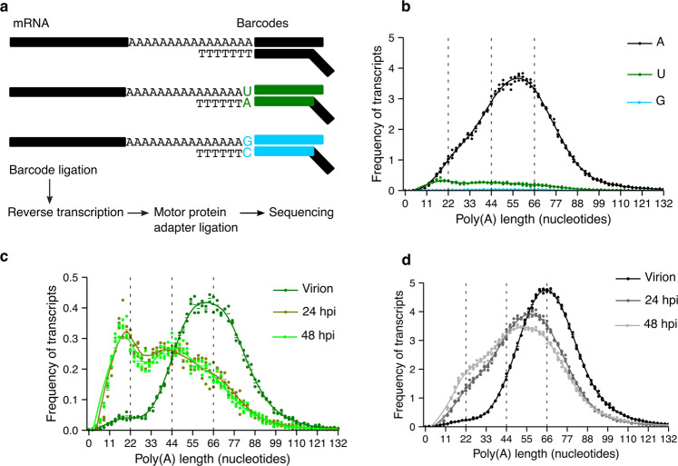
Fig. 1. MHV RNA is uridylated during infection.
a A diagram of the splint-ligation strategy used to capture RNA 3′-end modifications using barcoded oligos is shown. b The poly(A) tail length profile of MHV RNA from 17-CL1 infected cells 24 hours post-infection (hpi) is shown in black for tails without terminal modifications, in green for terminally uridylated tails, and in cyan for terminally guanylated tails. c The poly(A) tail length profile of terminally uridylated tails of the MHV virion RNA and MHV RNA from 17-CL1 infected cells at 24 and 48 hpi are indicated in dark green, olive, and light green, respectively. Uridylated reads represent 9.9%, 8.8%, and 8.7% of total reads in the virion RNA, the vRNA 24 hpi, and the vRNA 48 hpi, respectively. The profile of the uridylated transcripts at 24 hpi is the same as shown in b. d The poly(A) tail length profile of the MHV virion RNA and MHV RNA from 17-CL1 cells infected at 24 and 48 hpi are depicted in black, gray, and light gray, respectively. Dots represent the frequency of poly(A) tail length for each sample, and the horizontal bars show the mean frequency of poly(A) tail length per condition (n = 2 for the virion and n = 3 for 24 and 48 hpi).

