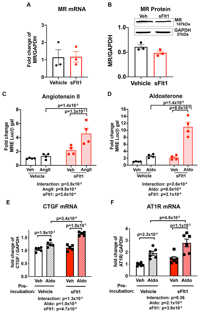
Figure 4. Transient exposure of smooth muscle cells (SMC) to sFlt1 in vitro enhances mineralocorticoid receptor (MR) transcriptional activity and target gene expression upon receptor stimulation.

Pac1 SMC biological replicates were exposed to 50 ng/mL sFlt1 for 24 hours and expression of MR; (A) mRNA and (B) protein was measured. Control n=3, sFlt1 n=3. Representative western blots of MR and GAPDH in SMC and quantification are shown. 24 hours after sFlt1 was removed, MR transcriptional reporter activity was measured in response to MR stimulation with (C) angiotensin II (AngII, all groups n=4 independent experiments, p values determined by 2-way ANOVA with Sidak post hoc or (D) aldosterone (Aldo, all groups n=4 independent experiments, p values determined by 2-way ANOVA with Sidak post hoc compared to vehicle treated controls. Aldo-stimulated mRNA expression of MR target genes; (E) connective tissue growth factor (CTGF, all groups n=5 independent experiments, p values determined by 2-way ANOVA with Sidak post hoc and, (F) AngII type 1 receptor (AT1R), all groups n=6 independent experiment, p values determined by 2-way ANOVA with Sidak post hoc.
