FIGURE 1.

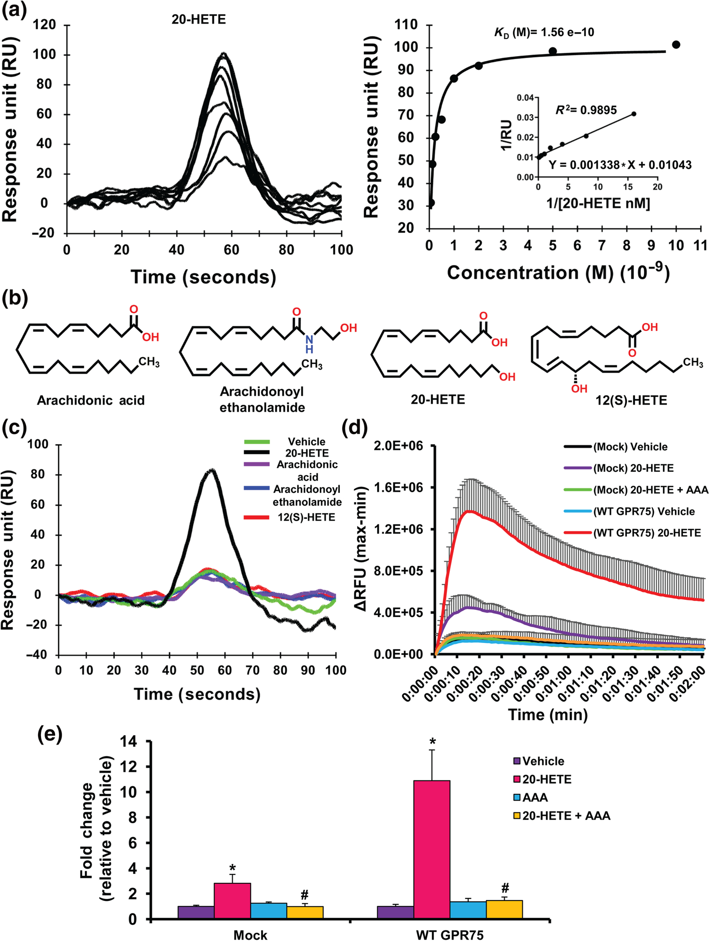
20-HETE binds to GPR75.(a) Surface plasmon resonance (SPR) analysis of 20-HETE-GPR75 binding, depicting the sensogram (normalized to vehicle (HBSS containing 1% ethanol) (left) of human recombinant GPR75 immobilized to the SPR sensor surface followed by 20-HETE injections (0.0625, 0.125, 0.25, 0.5, 1, 2, 5 and 10 nM) injected at a flow rate of 20 μl min−1 and affinity analysis (right). (b) Structures of arachidonic acid, arachidonoyl ethanolamide, 20-HETE and 12(S)-HETE. (c) SPR sensogram of human recombinant GPR75 immobilized to the SPR sensor surface followed by injections of vehicle (HBSS containing 1% ethanol), arachidonic acid, arachidonoyl ethanolamide, 12(S)-HETE and 20-HETE (1 nM) injected at a flow rate of 20 μl min−1; 20-HETE increases intracellular calcium (iCa2+) via GPR75. (d) FLIPR Calcium 6 assays of mock- or GPR75-transfected HTLA cells treated with vehicle (ethanol), 20-HETE (1 nM) and co-treatment of 20-HETE (1 nM) with AAA (1 nM), a 20-HETE receptor antagonist, showing post-treatment live calcium traces (2 min) or (e) peak treatment response expressed as fold change relative to vehicle-treated cells.Data shown are means ± SEM; n = 6.*P < 0.05, significantly different from vehicle (ethanol), #P < 0.05, significantly different from 20-HETE
