Fig. 2.
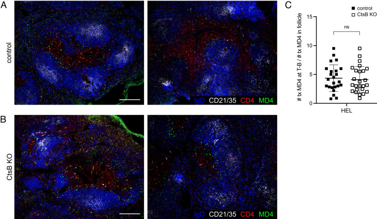
Follicular Exclusion of HEL-Engaged B Cells Is Unaffected by Ctsb Deficiency. (A and B) Immunofluorescence for MD4 GFP B cells (GFP, green) transferred into HEL-treated control (A) or Ctsb-deficient (B) mice stained to detect endogenous B cell follicles (IgD, blue; CD21/35, white) and the T cell zone (CD4, red). Sections were prepared one day after HEL treatment. Two example images are shown and are representative of multiple cross-sections from at least three mice of each type. (Scale bar, 200 µm.) (C) Quantification of proportion of MD4 GFP B cells at the T zone–follicle (T-B) interface one day after HEL treatment. Each data point represents an individual follicle (n = 24 control, n = 25 KO) from sections prepared from at least three mice of each genotype. Lines indicate means, and error bars represent SDs. Statistical significance was determined by unpaired t test. NS, not significant.

