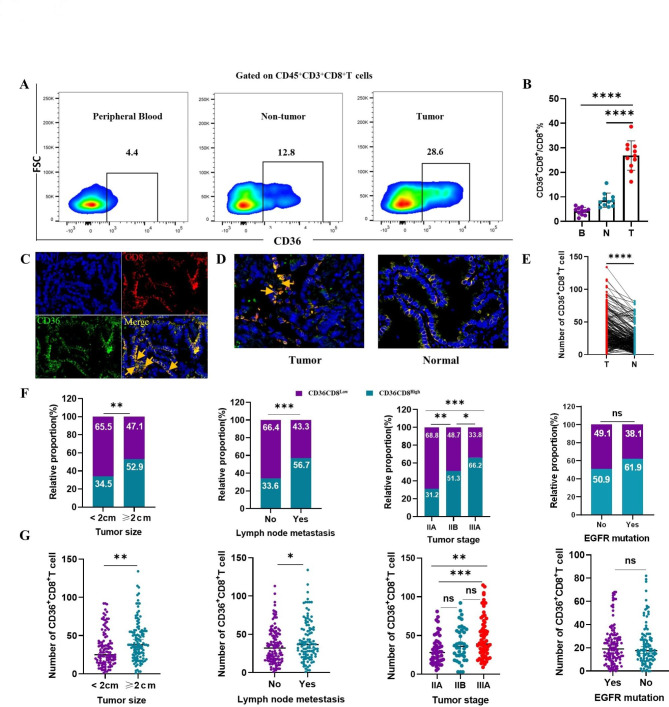
Fig. 2.
Accumulation of CD36+CD8+ T cells in NSCLC is correlated with disease progression. A, B. The representative flow cytometry and statistics analysis of CD36 expression on CD8+ T cells in matched PBMCs, non-tumor tissues and tumor from patients with NSCLC (n = 11, ****p < 0.0001 by paired t test). C. Representative images of the immunofluorescence staining with DAPI (blue), CD36 (green), CD8 (red), and merge (double positive) on NSCLC tissues. Scans were imaged at 200 magnifications. D, E. The proportion of CD36+CD8+ T cells is higher in tumor than that in non-tumor tissues (****p < 0.0001 by Student’ s t test). F, G. The number and proportion of CD36+CD8+ T cells was elevated in patients with larger tumor size (**p < 0.01), lymph node metastasis (***p < 0.0001) and advanced TNM stage (***p < 0.001). However, in patients with or without EGFR mutation, the number and proportion of CD36+CD8+ T cells was not statistically different (Chi square test and paired t test, respectively). B = blood, N = non-tumor, T = tumor

