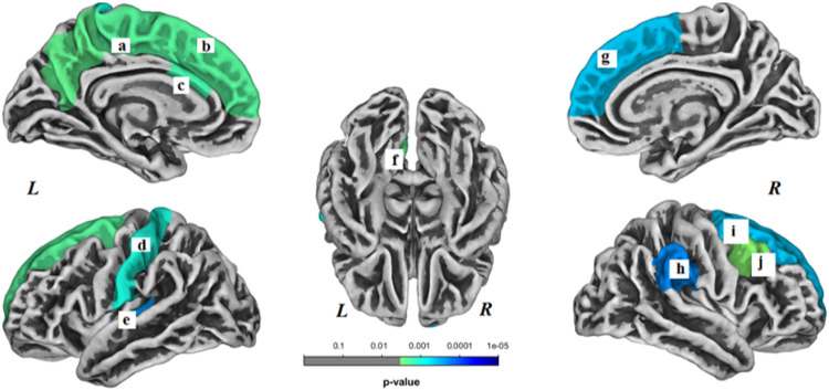
Figure 3.
Brain regions with decreased gyrification within the healthy control (HC) group. These regions included the following: (a) left paracentral gyrus, (b) left superior frontal gyrus, (c) left caudal anterior cingulate gyrus, (d) left postcentral gyrus, (e) left transverse temporal gyrus, (f) left precuneus, (g) right superior frontal gyrus, (h) right supramarginal gyrus, (i) right superior frontal gyrus, and (j) right caudal middle frontal gyrus. L- left hemisphere, R- right hemisphere. Results were Bonferroni correctedat significant level of 0.05.

