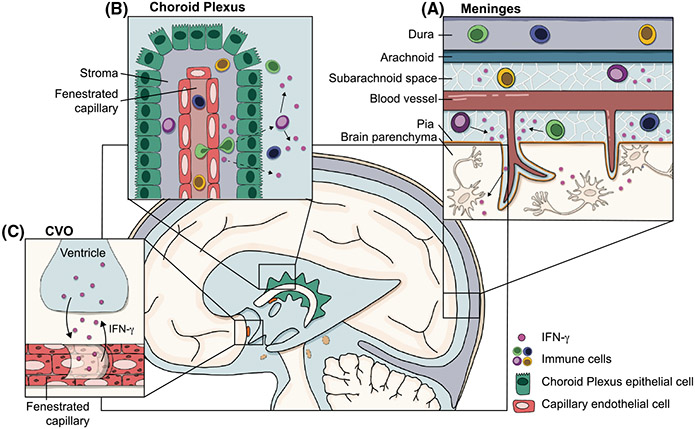
FIGURE 2.
Sources of IFN-γ in the CNS. Physiological levels of IFN-γ, as well as immune cells which can produce IFN-γ, are present in the CSF during steady state. Because it is hypothesized that the CSF connects with the interstitial fluid (ISF), which circulates throughout the brain parenchyma, we focus on the CSF as a potential carrier which may grant IFN-γ direct access to neurons. However, it is still unclear how IFN-γ gets into the CSF and ultimately into the parenchyma. A) The meninges contain an immunologically rich niche, including T cells, NK cells, and ILC1s, all of which can produce IFN-γ. Immune cells in the subarachnoid space and perivascular spaces may act as a source of physiological IFN-γ found in the CSF. B) The choroid plexus sits outside of the blood–brain barrier and instead acts as a blood–CSF barrier, where proteins and solutes can exit the fenestrated capillaries and enter the CSF. Immune cells (including IFN-γ producing T cells, NK cells, and ILC1s) have been identified in the choroid plexus stroma, but it is unclear whether they (or their secreted cytokines) can cross the epithelial layer of the choroid plexus to enter the CSF. C) Circumventricular organs (CVOs) are often referred to as the “windows of the brain,” as they also lack a classical blood–brain barrier. Early studies using HRP have demonstrated that HRP injected intraventricularly or intravenously is able to permeate the parenchyma, suggesting solutes can access the parenchyma from both the CSF and the blood. Though this has not been directly tested with IFN-γ, it may be a potential route for IFN-γ to gain access to the parenchyma. Additionally, studies have demonstrated that cytokines can exit the fenestrated capillaries and act directly on the cells of the CVOs. However, it is still unclear whether cytokines can enter the CSF through CVOs, or whether cytokines have direct access to neurons within CVOs

