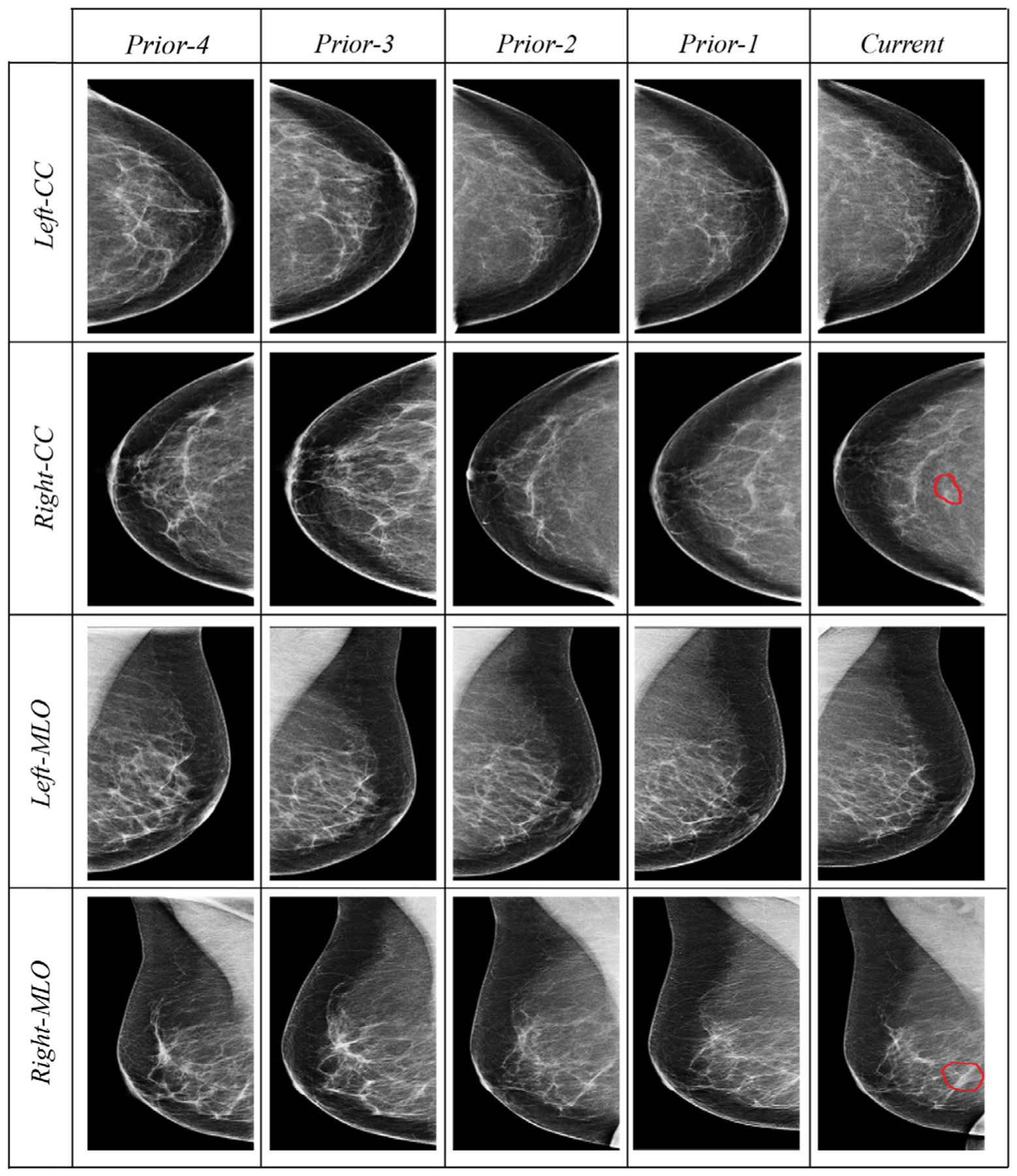
Fig. 1.

Examples of four longitudinal prior mammogram examinations (16 mammogram images) belonging to a 54-year-old woman diagnosed with nuclear grade 2 ductal carcinoma in situ manifest as microcalcifications in the right breast (red contours outlining the tumor region in the “current” column). Prior-1 through prior-4 are respectively captured at 1, 2, 3, and 4 year(s) earlier the cancer diagnosis at the current mammogram. Each prior includes four images: two projections (mediolateral oblique, MLO, and craniocaudal, CC) of each of the right and left breasts. (For interpretation of the references to colour in this figure legend, the reader is referred to the web version of this article.)
