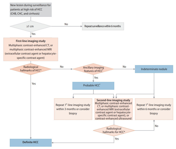
Figure 1.
Diagnostic algorithm of HCC. HCC, hepatocellular carcinoma; CHB, chronic hepatitis B; CHC, chronic hepatitis C; CT, computed tomography; MRI, magnetic resonance imaging; APHE, arterial phase hyperenhancement; US, ultrasonography. *The radiological hallmarks for diagnosing “definite” HCC on multiphasic contrast-enhanced CT or MRI are APHE with washout appearance in the portal venous, delayed, or hepatobiliary phase. These criteria should be applied only to a lesion that does not show either marked T2 hyperintensity or targetoid appearance on diffusion-weighted images or contrast-enhanced images. For a second-line imaging modality, contrast-enhanced US (blood-pool contrast agent or Kupffer cell-specific contrast agent) for a “definite” diagnosis of HCC is APHE with mild and late (≥60 seconds) washout. These criteria should be applied only to a lesion that does not show either rim or peripheral globular enhancement in the arterial phase. †For diagnosis of “probable” HCC, ancillary imaging features are applied as follows. There are two categories of ancillary imaging features, those favoring malignancy in general (mild-to-moderate T2 hyperintensity, restricted diffusion, threshold growth) and those favoring HCC in particular (enhancing or non-enhancing capsule, mosaic architecture, nodule-in-nodule appearance, fat or blood products in the mass). For nodules without APHE, “probable” HCC can be assigned only when the lesion fulfills at least one item from each of the two categories of ancillary imaging features. For nodules with APHE but without washout appearance, “probable” HCC can be assigned when the lesion fulfills at least one of the aforementioned ancillary imaging features. Adopted from 2022 KLCA-NCC HCC guidelines [1].

