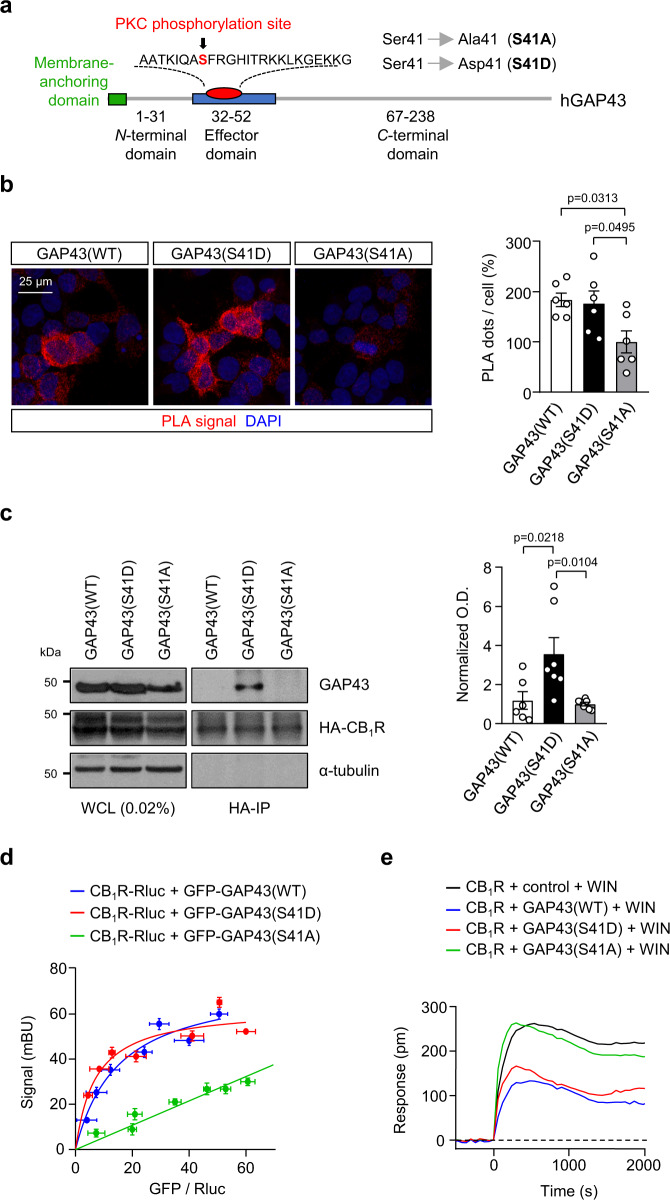
Fig. 2. Phosphorylation of GAP43 at S41 facilitates its interaction with CB1R.
a Scheme of the mutant constructs aimed to modify GAP43 activation state. b PLA for CB1R and GAP43 was performed with anti-c-myc and anti-GFP antibodies in HEK293T cells transfected with CB1R-myc plus GFP-GAP43(WT), GFP-GAP43(S41D) or GFP-GAP43(S41A). Left, Representative confocal microscopy images show CB1R-GAP43 complexes appearing as red dots. Cell nuclei were stained with DAPI (blue). Right, Quantification of PLA-positive dots per GFP-transfected cell. Values of GFP-GAP43(S41A) were set at 100% (means ± SEM; n = 6 independent experiments; one-way ANOVA with Tukey’s multiple comparisons test). c Left, Co-immunoprecipitation experiments in HEK293T cells co-transfected with HA-tagged CB1R and GAP43(WT), GAP43(S41D), GAP43(S41A). Whole-cell lysates (WCL) are shown. Right, Quantification of optical density (O.D.) values of co-immunoprecipitated GAP43 relative to those of HA-CB1R are shown. Values of GFP-GAP43(S41A) were set at 1 (means ± SEM; GAP43(WT) n = 6 independent experiments, GAP43(S41D) n = 7 independent experiments, GAP43(S41A) n = 7 independent experiments; one-way ANOVA with Tukey’s multiple comparisons test). d BRET saturation experiments in HEK293T cells expressing CB1R-RLuc and increasing amounts of GFP-GAP43(WT), GFP-GAP43(S41D) or GFP-GAP43(S41A). BRET is expressed as milli-BRET units (mBU) (means ± SEM; n = 3 independent experiments). e DMR assays in HEK293T cells transfected with CB1R plus GAP43(WT), GAP43(S41D), GAP43(S41A) or a control empty vector, and exposed to 100 nM WIN-55,212–2 (WIN). A representative experiment is shown (n = 3 independent experiments). Source data are provided as a Source data file.

