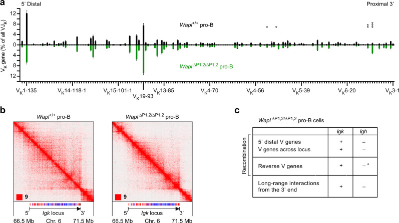
Fig. 4. VK-JK recombination at the Igk locus in Waplhigh and Wapllow pro-B cells.
a VK gene recombination analysis of ex vivo sorted Wapl+/+ (Wapllow; black) and Wapl∆P1,2/∆P1,2 (Waplhigh; green) pro-B cells, as determined by VDJ-seq experiments. The recombination frequency of each VK gene is indicated as a percentage of all VK-JK rearrangement events and is shown as a mean value with SEM based on six independent VDJ-seq experiments for each pro-B cell type. The different VK genes (horizontal axis) are aligned according to their position in the Igk locus25 (Supplementary Data 1a). Statistical data were analyzed by multiple t-tests (unpaired and two-tailed) with Holm–Sidak correction; *P < 0.05, **P < 0.01, ***P < 0.001. b Hi-C contact matrices of the Igk region on chromosome 6 based on published Hi-C data of short-term cultured Wapl+/+ and Wapl∆P1,2/∆P1,2 pro-B cells20. The orientation and annotation of the Igk locus are shown. The resolution of the Hi-C data was 6.7 and 7.25 kb for the Wapl+/+ and Wapl∆P1,2/∆P1,2 pro-B cells, respectively. c Differences in V gene recombination and long-range interactions between the Igh and Igk locus in Wapl∆P1,2/∆P1,2 pro-B cells, based on the data shown in Fig. 4 and published data20. The loss of recombination of VH genes upon their inversion (indicated by an asterisk) in the Igh locus was analyzed in IghV8-8-inv/V8-8-inv and Igh∆890/∆890 pro-B cells20 and upon inversion of the entire VH gene cluster in v-Abl immortalized pro-B cells32. Source data are provided in the Source Data file.

