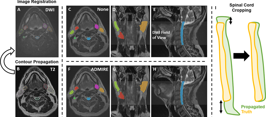
Figure 1.
Study workflow. Contours are propagated from the moving image (B, T2-weighted image [T2]) to the fixed image (A, diffusion-weighted image [DWI]) for each registration method. C-E and F-H show propagated and ground-truth structures for no registration (labeled as “None”) and ADMIRE registration approaches, respectively. The spinal cord was also cropped so that the height of the propagated segmentation and ground-truth segmentation were equal (I).

