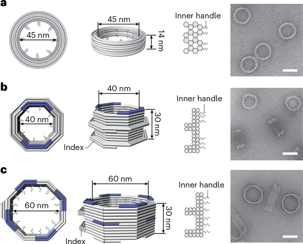
Fig. 1 |. Design and assembly of DNA-origami channels as mimics of the NPC scaffold.

a, A DNA cylinder with an inner diameter of 45 nm, dubbed 45-nm channel. b, An open-ended DNA octagonal prism with an inner diameter of 40 nm, dubbed 40-nm channel. c, An open-ended DNA octagonal prism with an inner diameter of 60 nm, dubbed 60-nm channel. The 40-nm (b) and 60-nm (c) channels are designed with protruded regions on the side, or indexes, to mark the polarity of the channel, as well as shape-complementary interfaces (blue) at the top and bottom to facilitate vertical dimerization. Shown from left to right are cartoon models of the DNA-origami channels (in right, perspective and cross-section views) and representative negative-stain electron micrographs. All negative-stain EM experiments were repeated three times with similar results. The gray curls represent ssDNA extensions (handles) for organizing desired nups. Scale bar, 50 nm.
