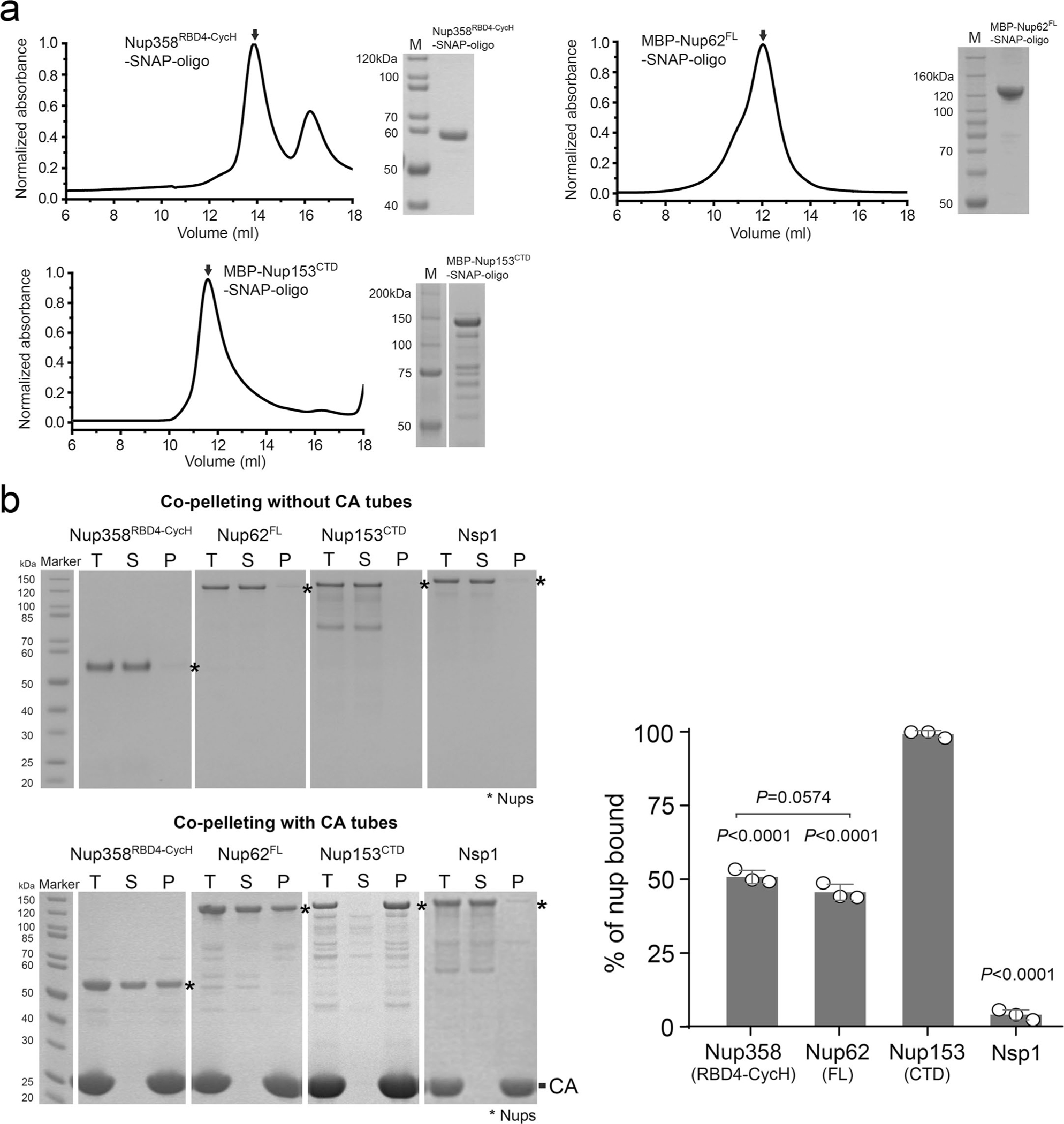
Extended Data Fig. 3 |. Nup purification and CA nanotube co-pelleting assays.

a, Size exclusion chromatography analysis showing that the DNA-conjugated of Nup358RBD4-CycH, MBP-Nup62FL, and MBP-Nup153CTD are mainly monodispersed monomers in solution. Right panels: SDS-PAGE analysis of protein purity. The experiment was repeated three times with similar results. b, Left: co-pelleting assay of purified nups using A14C/E45C disulfide crosslinked CA nanotubes. Soluble (S) and pellet (P) denote fractions of co-pelleting assays. Total (T) denotes the nup-CA tube mixture before fractionation. Right: percentages of various nups bound to CA tubes in the co-pelleting assays in PBS buffer. Data are plotted as mean±SD of three independent experiments (n = 3), with individual data points shown as black circles. Differences were determined by one-way ANOVA and Tukey’s multiple comparisons test. Unless marked in the graph, comparisons were made with MBP-Nup153CTD. Full statistical results are provided in source data.
